top of page
13.png

Antibody Profiling Technology

Antibody profiling refers to the process of simultaneously detecting a large number of antigen-antibody interactions. Technologies for antibody profiling typically involve testing a blood or a monoclonal antibody sample against a large library of antigens.


The antibodies in the specimen bind to their target antigens, and the binding is then analyzed to determine the presence and levels of each antigen-reactive antibody. This information can be used to understand disease mechanisms, develop better diagnostic tests, treatments, and vaccines.

There are several methods for performing antibody profiling, including microarray and other molecular display-based methods. However, previous methods have been limited by their library content, slow turnaround time, and/or high cost. MIPSA is a powerful new molecular display-based method that uses self-assembly to provide the greatest flexibility in library content and the fastest turnaround time, all at significantly lower cost.


How does Infinity Bio read antibody reactivities?

​

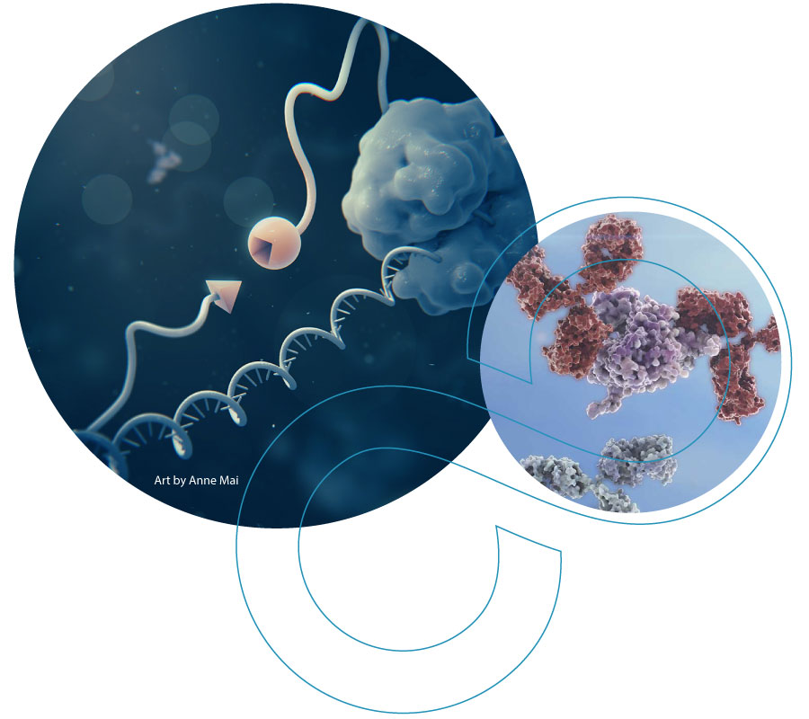
MIPSA employs self-assembly to create a library of in-solution DNA-barcoded antigens. Once bound by an antibody and then pulled down, sequencing of the DNA barcodes maps the antibody reactivities repertoire.

​

​

​

​

​

​

​

​

​

​

​

​

​

​

​

​

​

​

​


Antibody profiling with MIPSA involves three key steps:

Screen Shot 2024-01-11 at 4.19.27 PM.png
Screen Shot 2024-01-11 at 4.23.41 PM.png

1. Library creation

GENE SYNTHESIS AND CLONING
MIPSA is used to express

(i) libraries of cloned Open Reading Frames (ORFs) that encode full-length proteins

and/or

(ii) synthetic DNA oligonucleotide libraries that encode peptides designed to tile across proteins.

​

Both types of libraries can be used for antibody profiling in the same reaction.

Full-Length

Screen Shot 2023-11-01 at 10.15.09 AM.png

(i)

Screen Shot 2023-08-11 at 11.34.34 AM.png

Tiled Peptide

Screen Shot 2023-08-11 at 11.34.34 AM.png

(ii)

SELF-ASSEMBLY: ATTACHING THE DNA BARCODE TO THE PROTEIN
The self-assembly step of MIPSA technology results in the covalent attachment of a DNA barcode to the protein or peptide library member via the HaloTag system.
This process is accomplished in three steps:
A) Creation of the barcoded RNA library,
B) Reverse transcription, and
C) Translation of the HaloTagged antigen library.

A. Creation of the barcoded RNA library

The MIPSA process begins with engineered vectors that include the following key elements:

  • an RNA polymerase transcriptional start site,

  • a barcode sequence flanked by universal polymerase chain reaction (PCR) primer binding sites,

  • a translation start site,

  • an N-terminal HaloTag protein,

  • and the sequence encoding the library member (target antigen).

​

The in vitro transcribed RNA consists of the unique barcode, the sequence encoding the HaloTag protein and the sequence encoding the library member.

Screen Shot 2023-08-11 at 11.34.39 AM.png

B. Reverse transcription

Reverse transcriptase synthesizes DNA by using a reverse transcription (RT) primer that is conjugated with a HaloLigand.

Screen Shot 2023-09-14 at 2.41.10 PM.png
Screen Shot 2023-08-11 at 11.34.39 AM.png

HaloLigand

C. Translation of HaloTagged antigen library

The HaloTag protein domain is translated first, followed by the translation of the antigen protein or peptide library member.


The HaloTag protein domain has a cleft which covalently attaches to the HaloLigand, thereby linking the DNA barcode and the antigen library protein or peptide in a process of "self assembly". 

​

5.png

HaloLigand

Screen Shot 2023-08-11 at 11.34.39 AM.png
4.png

DNA-BARCODED PEPTIDES OR FULL-LENGTH PROTEIN LIBRARIES

​

The resulting MIPSA library consists of a large number of antigens, each associated with a unique set of DNA barcodes. 

An antibody-containing sample is incubated with an individual library or a combination of libraries, allowing the antibodies to find and bind their target antigen library members. 

image.png

DNA-barcoded Full Length Protein 

and/or

image.png
image.png
image.png

DNA-barcoded peptide antigens

2. Antibody-antigen interaction 

LINEAR & CONFORMATIONAL EPITOPES
An antibody epitope, also known as an antigenic determinant, is the part of the antigen that is specifically recognized by an antibody.


Conformational epitopes are formed by the 3D folding of the antigen molecule, frequently involving discontinuous (non-contiguous) amino acid sequences that are brought together in space. Detection of antibodies targeting conformational epitopes typically requires larger fragments or full-length proteins.


Linear epitopes tend to lack significant 3D structure and are determined by the primary sequence of contiguous amino acids. Detection of antibodies targeting linear epitopes is often accomplished using peptides. 

image.png
image.png

3. Decoding of information

ANTIBODY-BOUND MIPSA LIBRARY MEMBERS ARE CAPTURED USING MAGNETIC BEADS
After antibody-antigen complexes are formed in solution, magnetic beads are then used to capture the antibodies along with each bound MIPSA library member and its attached cDNA barcode.

image.png
image.png
image.png
image.png

DNA SEQUENCING AND ANALYSIS
After unbound MIPSA library members are washed away, PCR is used to amplify the MIPSA barcodes. Many samples can be analyzed together on a sequencing instrument by using sample-specific barcoded primers for the PCR step. High throughput DNA sequencing is used to count the number of MIPSA library member molecules captured in each sample. Proprietary software pipelines convert the sequencing count data into interpretable assay results.


Screen Shot 2023-09-12 at 2.21.46 PM.png

EXAMPLE SAMPLE DATA: SARS-COV-2

Antibody-peptide binding data can be mapped onto viral protein structures, as shown here for one patient after recovering from a SARS-CoV-2 infection, for example.

Screen Shot 2023-10-03 at 9.21.19 AM.png

Ready to get started?

bottom of page